SEMKUR - Imaging
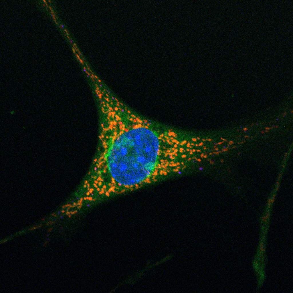
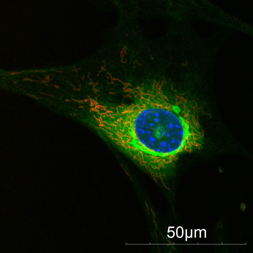
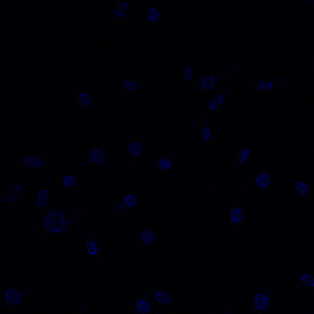
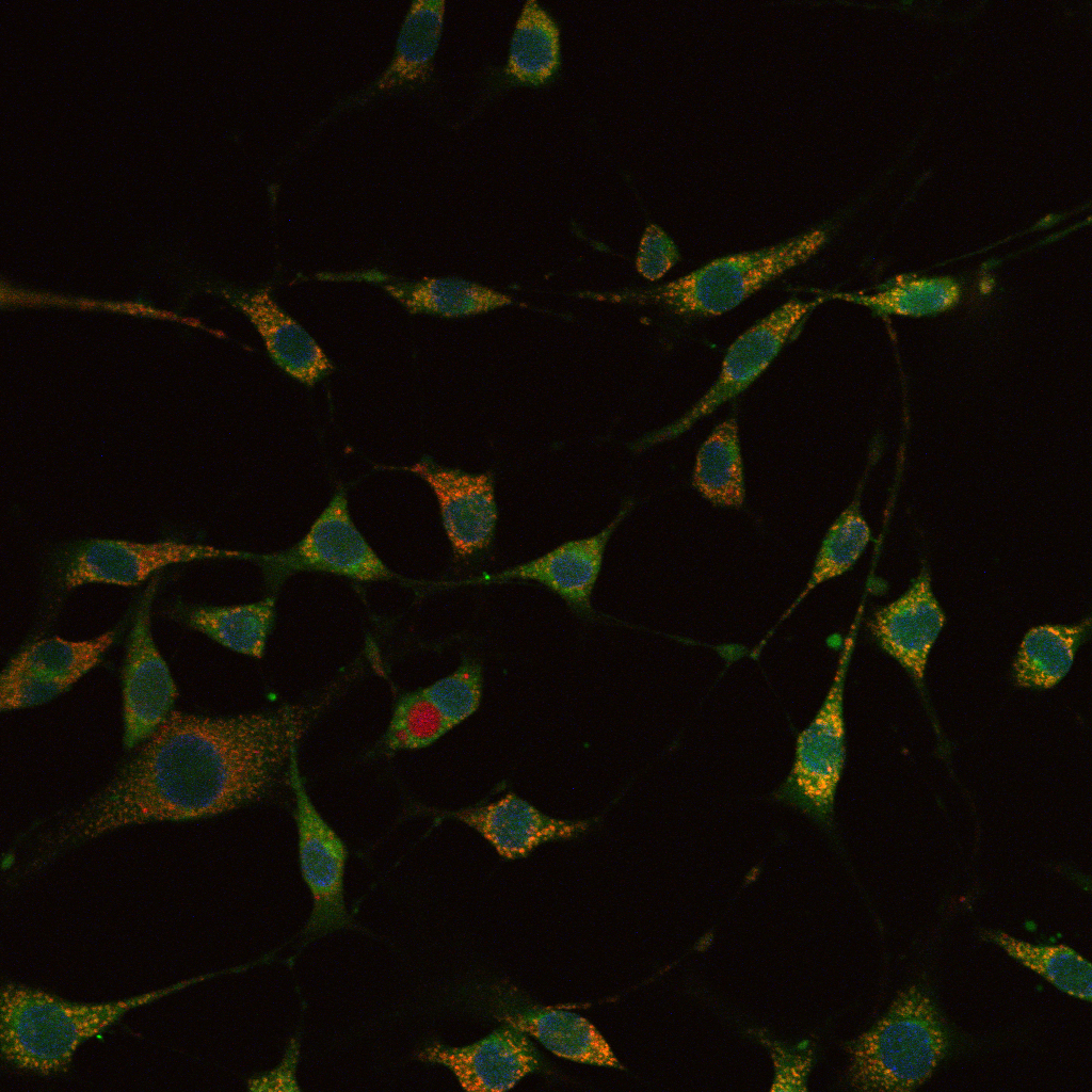
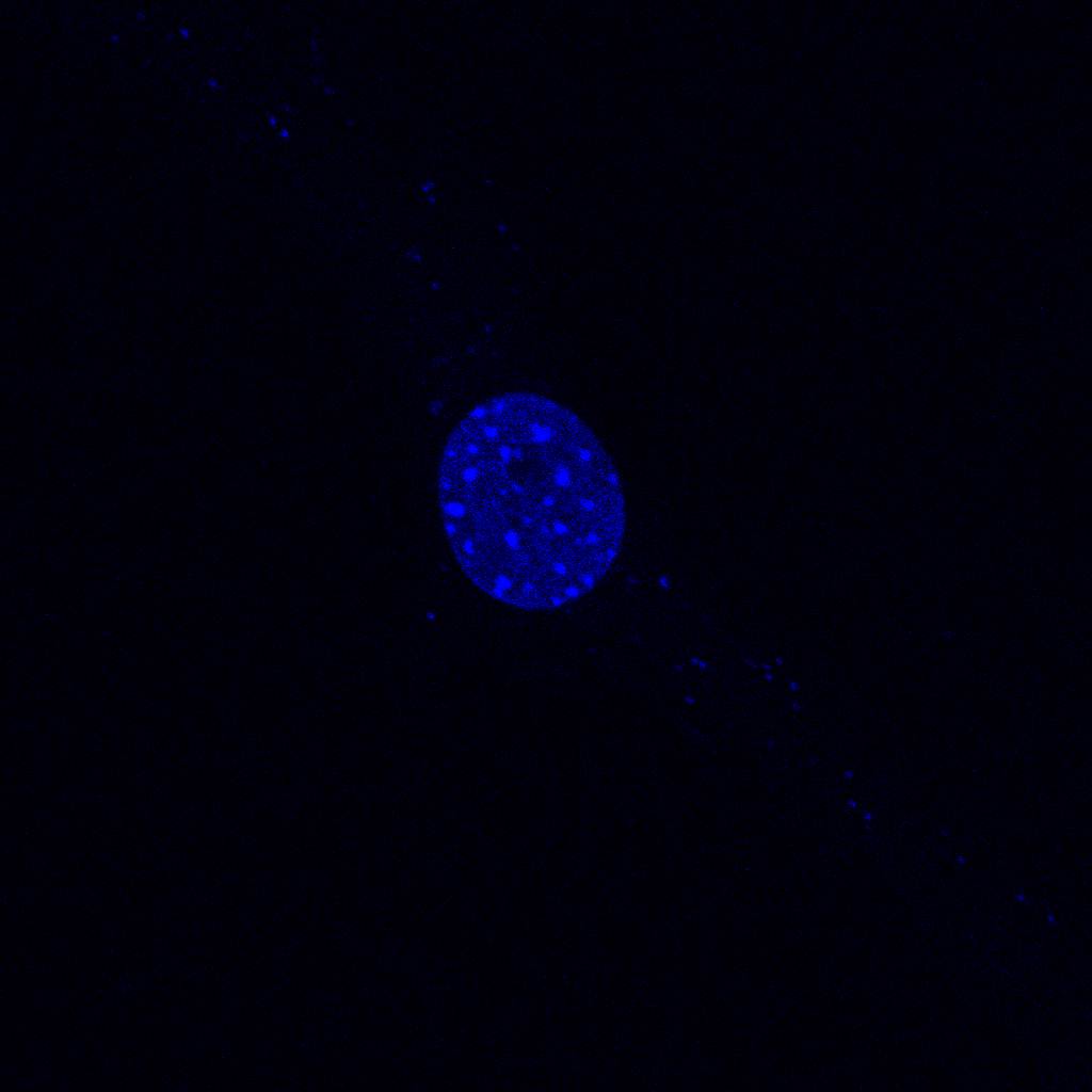
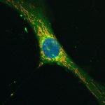
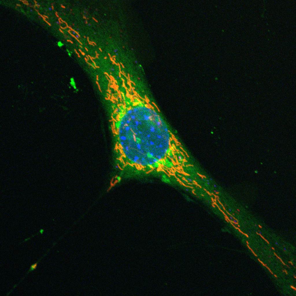
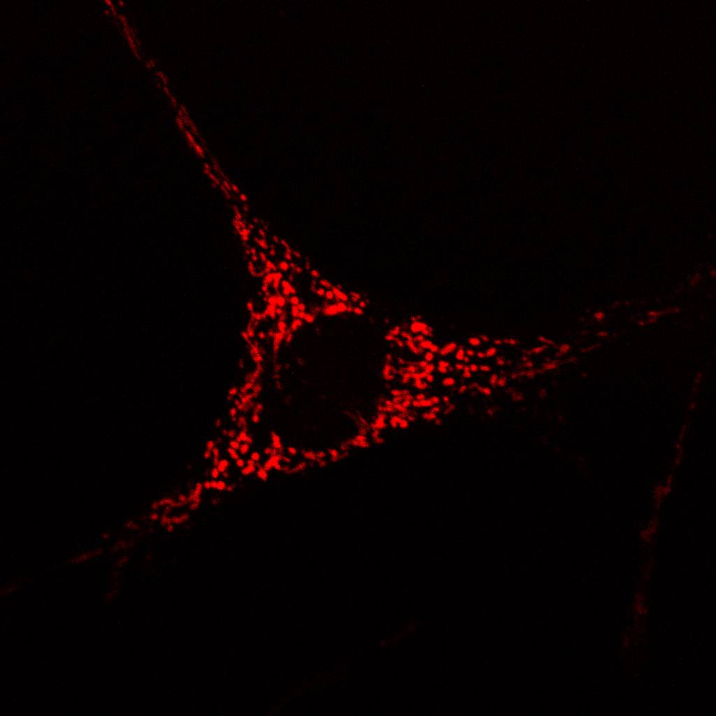
NIH/3T3 cells are plated with SEMKUR-Imaging at a 20 micromolar concentration and incubated with CO2 for 24 hours in complete media on a MatTek Dish. The media was then washed off and replaced with Hoechst, blue nuclear stain at a concentration of 1 microgram/ml. TMRM mitochondrial red stain was introduced at a concentration of 200nm while SEMKUR-Imaging green stain was used at a concentration of 20 micromolar. These stains were combined to show colocalization of the SEMKUR stain in the mitochondria.
Single Cell Co-Localization Study
Click an image to view larger-
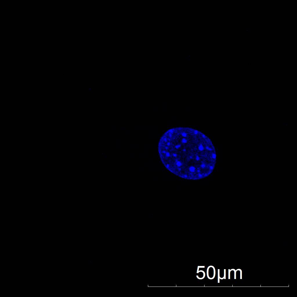
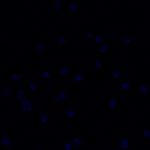
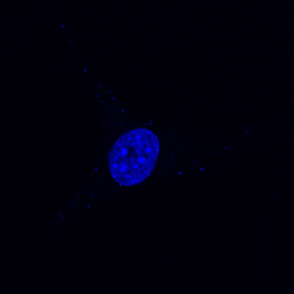
- Hoechst probe
-
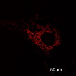
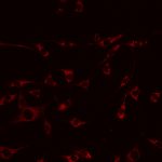
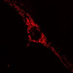
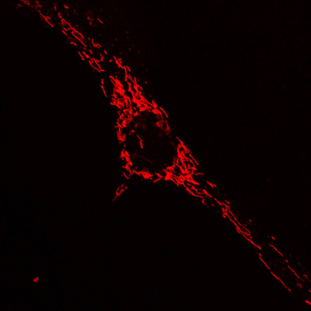
- Mitochondrial probe

-
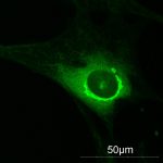
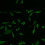
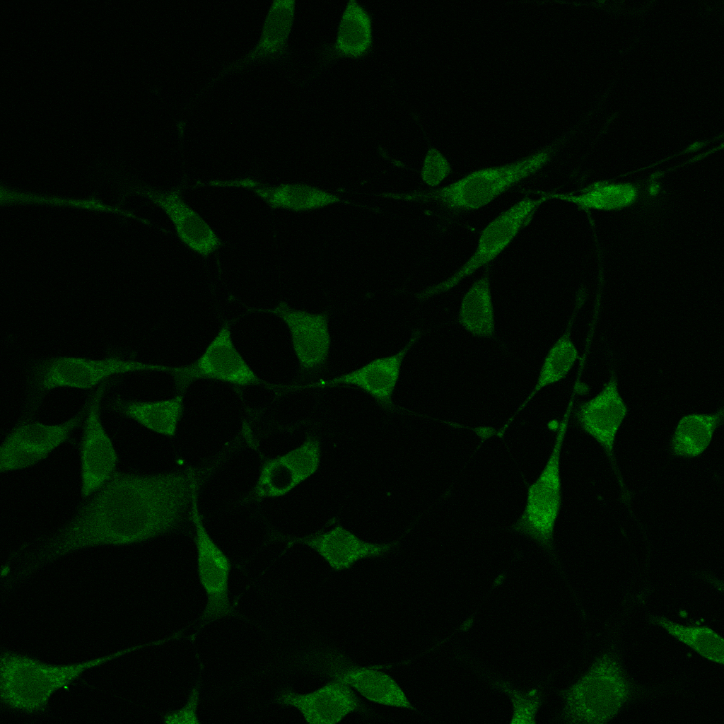
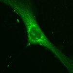
- SEMKUR-imaging probe

-
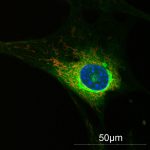
- All probes
Multi-Cell Co-Localization Using 3T3 Cells
Click an image to view larger-
- Hoechst probe
-
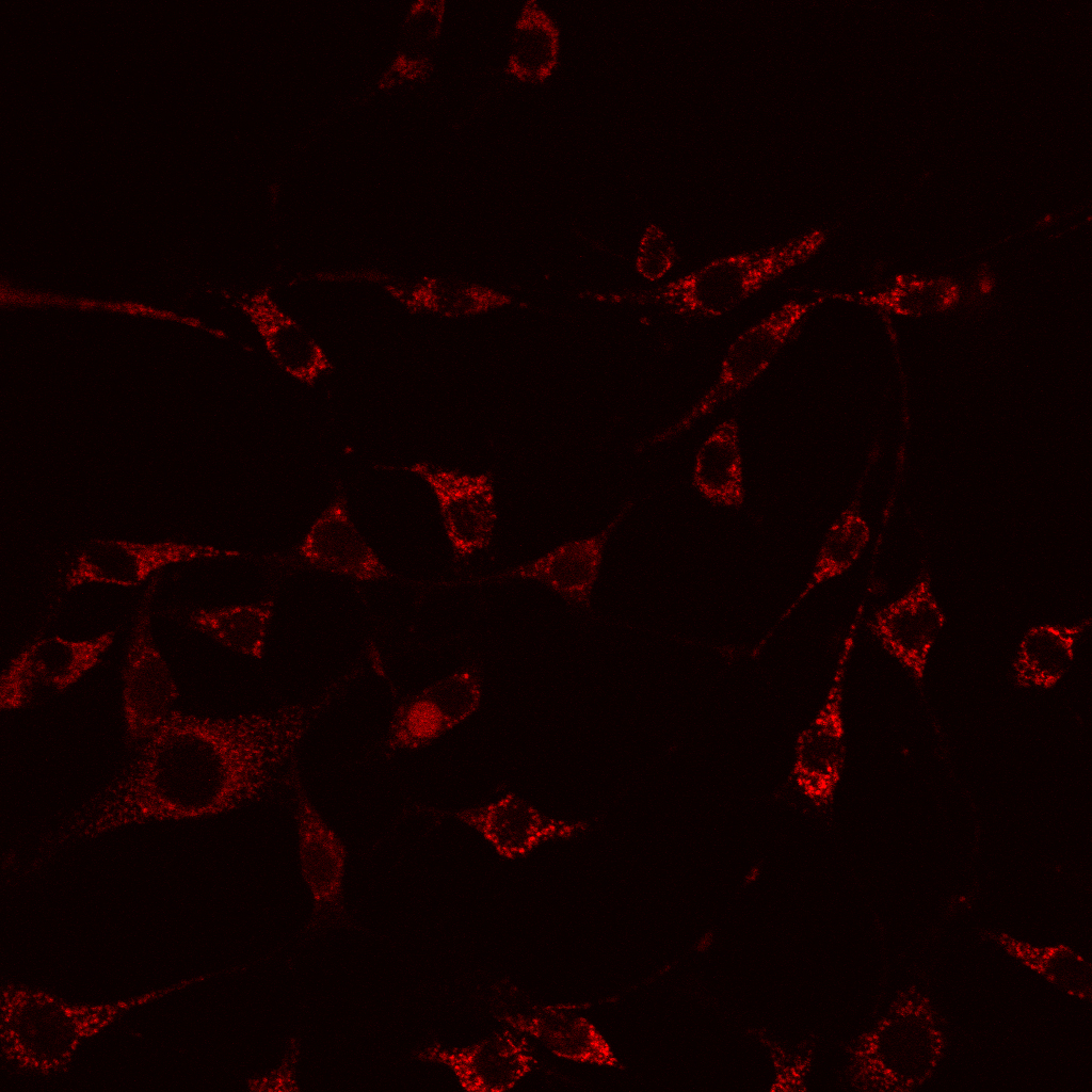
- Mitochondrial probe
-
- SEMKUR-imaging probe
-
- All probes
Co-Localization Study
Click an image to view larger-
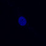
- Hoechst probe
-
- Mitochondrial probe
-
- SEMKUR-imaging probe

-
- All probes
Co-Localization Study
Click an image to view larger-
- Hoechst probe
-
- Mitochondrial probe
-

- SEMKUR-imaging probe

-

- All probes